Dendrimer-based nanosystems, described in the journal Advanced Materials, offer a promising nanotechnological platform for the development of multimodal imaging probes for biomedical applications. This result is the fruit of the work of scientists from the Centre interdisciplinaire de nanoscience de Marseille (CNRS/Aix Marseille Université) and the Centre de résonance magnétique biologique et médicale (CNRS/Aix Marseille Université).
Reading time: 3 minutes
|
Key facts to remember:
|
Using medical imaging to diagnose and assess treatment efficacy
Biomedical imaging enables the non-invasive diagnosis and monitoring of tumors, as well as the evaluation of treatment efficacy. Magnetic resonance imaging (MRI) is one of the most commonly used techniques for diagnosis and follow-up, while the use of intraoperative near-infrared fluorescence spectrometry (NIRF) is constantly progressing. The former provides anatomical, functional and metabolic information, and often requires the use of gadolinium (Gd3+)-based contrast agents to enhance contrast between tumors and surrounding tissue. Fluorescence imaging, with its high sensitivity but more limited tissue penetration, is increasingly used in the operating room to guide surgery. It requires the use of specific near-infrared dyes (NIRF).
Enhancing measurement sensitivity and specificity
Combining these two techniques should make it possible to increase the sensitivity and specificity of measurements. To achieve this, scientists from the Centre interdisciplinaire de nanoscience de Marseille and the Centre de résonance magnétique biologique et médicale have devised nanosystems that can act as both contrast agents for MRI and dyes for NIRF. They have chosen amphiphilic dendrimers, tree-like molecules whose multiple endings can bind Gd3+ ions. In the core of nanoparticles assembled from these dendrimers, they then encapsulated a specific NIRF dye, enabling them to combine MRI and NIRF measurements. In this way, they obtained high-quality "bimodal" images of a pancreatic tumor.
Double the contrast and signal intensity
These dendrimers showed a twofold improvement in contrast in tumors compared with adjacent healthy muscle, and a twofold increase in fluorescence signal intensity. What's more, these systems were shown to be effective even at Gd3+ concentrations corresponding to 1/10 of the standard clinical dose, which could prevent potential risks of Gd3+-related toxicity.
These dendrimer-based nanosystems, described in the journal Advanced Materials, offer a promising nanotechnological platform for the development of multimodal imaging probes for biomedical applications.
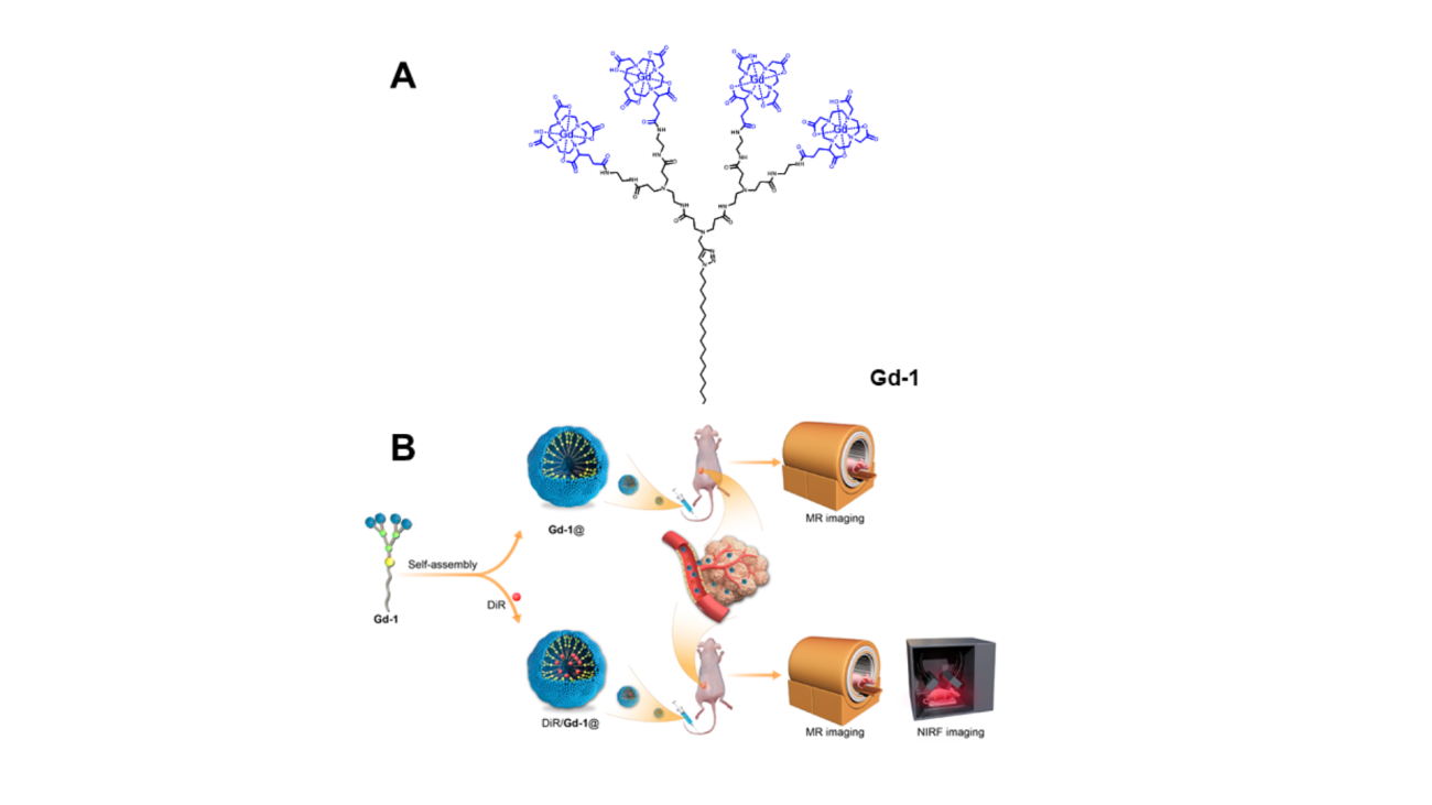
Nanosystèmes de dendrimères auto-assemblés pour l’IRM et l’imagerie multimodale des tumeurs. (A) Structures chimiques du dendrimère amphiphile Gd-1. (B) Illustration schématique des nanosystèmes de dendrimères auto-assemblés en tant qu’agent IRM (Gd-1@) et agent d’imagerie bimodale MR/NIRF (DiR/Gd-1@) des tumeurs. © Ling Peng
Contact à ajouter
Informations complémentaires
Contact à ajouter
Reference : Ding L, Lyu Z, Perles-Barbacaru TA, Huang AY, Lian B, Jiang Y, Roussel T, Galanakou C, Giorgio S, Kao CL, Liu X, Iovanna J, Bernard M, Viola A, Peng L. Modular Self-Assembling Dendrimer Nanosystems for Magnetic Resonance and Multimodality Imaging of Tumors. Adv Mater. 2024 Feb;36(7):e2308262.
Article published March 28, 2024.
Discover also


Share on social media or