Endocytosis is a mechanism for transporting molecules into the cell. This mechanism is fundamental to the functioning of neurons. In an article published in the journal Science, scientists from the Institute of Neurophysiopathology (amU/CNRS) and the Institute of Myology (AP-HP/CEA/CNRS/Inserm/Sorbonne University) describe new structures, which they call "clearing", along the initial segment of the axon and which enable endocytosis "on demand" under the effect of physiological stimuli.
Reading time: 4 minutes
|
Key facts to remember:
|
Endocytosis along the axon
Endocytosis in neurons is primarily mediated by the assembly of wells coated with clathrin, a structural protein that forms the envelope of certain vesicles and enables invagination of the plasma membrane and formation of endocytosing vesicles. In neurons, these wells are numerous in the cell body and dendrites, as well as around the active zone in presynapses, the structure of the neuron that enables nerve communication. In contrast, little is known about the nanoscopic organization or presence of clathrin-mediated endocytosis along the axon, the part of the neuron that transmits electrical signals to target cells, particularly in the initial segment of the axon, a specialized compartment located at the beginning of the axon. It was conventionally assumed that endocytosis along the axon was minimal, a hypothesis supported by the presence of a dense complex of spectrins, ankyrins and membrane proteins anchored at the initial segment, with the presence of a periodic scaffold of actin rings linked by spectrins along the plasma membrane.
Evidence of "clearings" that allow clathrin wells to form along the axon
In a paper published in the journal Science, scientists have combined super-resolution and electron microscopy to reveal the nano-architecture of clathrin wells at the initial segment and their endocytosis activity. In addition to the periodic scaffolding, spectrins form well-defined circular exclusion zones with actin, which they termed "clearings", allowing clathrin wells to form at their center, in contact with the plasma membrane. By genetically manipulating neuron cultures or treating them with substances targeting actin or the spectrin network, they discovered that the dense spectrin network limits well formation by defining these clearings. The clathrin wells contained in the initial segment clearings are unusually stable, restricting endocytosis to the initial segment of resting neurons. These immobilized wells can be "unlocked" and endocytosis triggered at the initial segment by stimulation with N-methyl-D-aspartate (NMDA). This triggering of endocytosis is mediated by the polymerization of branching actin "nests" within the spectrin clearings around the clathrin-coated pits, which help to detach the clathrin wells from the plasma membrane.
Endocytosis is regulated by clearing in the axon's initial segment
This nanoscale study reveals that endocytosis at the initial axon segment is regulated at two levels. Firstly, the dense spectrin mesh limits the formation of clathrin pits within circular clearings where they are stabilized, resulting in low steady-state endocytosis activity. Secondly, endocytosis from these pre-formed wells can be triggered by physiological signals. These two levels of regulation explain how endocytosis is regulated within the stable scaffold of the mature axon, enabling its structural rearrangement and the regulation of neuronal excitability.
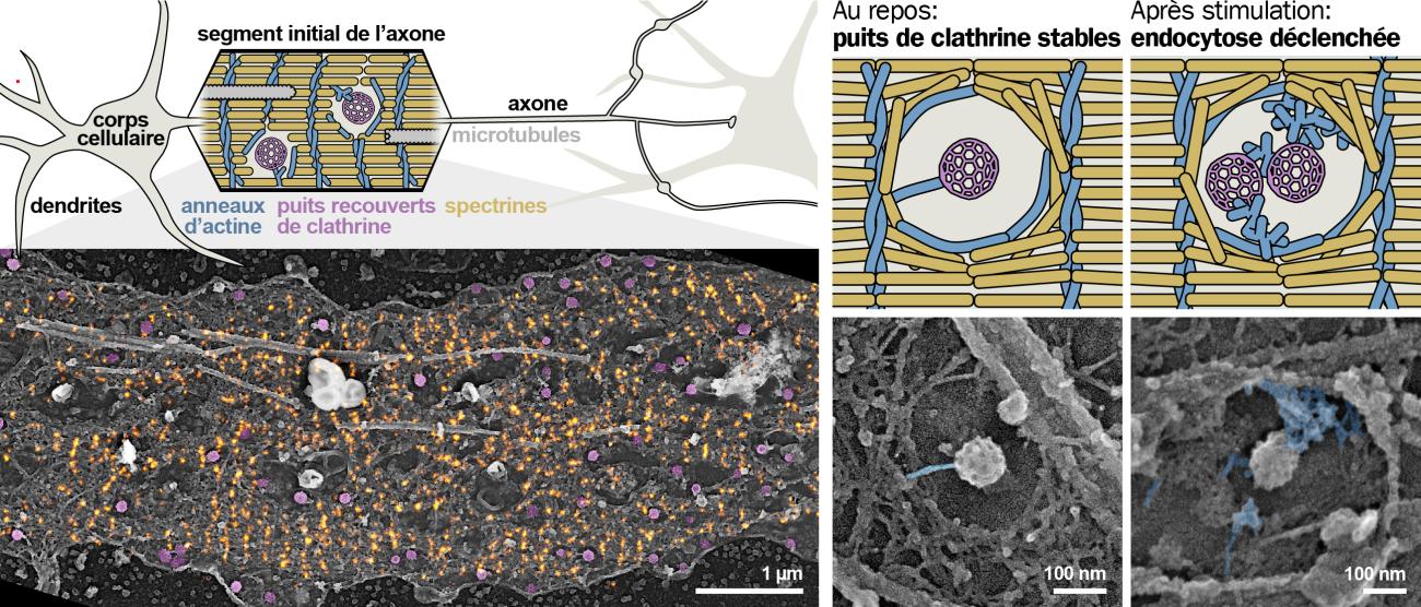
Illustration: Nanoscopy of clathrin-coated wells and endocytosis at the initial segment of the axon. Along the initial segment of the axon (top left), clathrin-coated pits (in magenta on the STORM/electron microscopy PREM super-resolution correlative view, bottom left) form in the clearings of the periodic spectrin scaffold (in orange). Endocytosis of these stable wells within the clearings is triggered after NMDA stimulation via polymerisation of branched actin nests (blue in the electron microscopy images and diagrams, right).
Contact à ajouter
Informations complémentaires
Article published on September 11, 2024.
Reference : F. Wernert, S. Babu Moparthi*, F. Pelletier, J. Lainé, G. Moulay, E. Simons, F. Boroni-Rueda, N. Jullien, S. Benkhelifa-Ziyat, M.J.Papandréou, C. Leterrier, S. Vassilopoulos, The actin-spectrin submembrane scaffold restricts endocytosis along proximal axons. Science 385, eado2032 (2024).
Picture credit : © Christophe Leterrier and Stéphane Vassilopoulos
Discover also


Share on social media or