

Are you ready for an unusual and exciting journey into the intricacies of the human brain? Meet Vincent Di-Marino, co-author of the Anatomical and Photographic Atlas of the Brain, which offers a complete photographic panorama of this fabulous organ.
Reading time: 4 minutes
You've already devoted several books to different parts of the human anatomy. Why did you turn your attention to the brain this time?
When we embark on a career as long as medicine, we hope to learn all the facets of human anatomy and eventually discover a passion for one of them. For me, it was neuroanatomy, and more particularly the part devoted to the brain, taught magnificently in Marseille by Prof. Paul Bourret, who patiently drew the nerve structures and their connecting bundles on the blackboard. When I became a hospital extern, I was lucky enough to be assigned to the neurology department at La Timone, where I attended consultations with prestigious neurologists who were able to make precise diagnoses based on clinical examination alone and their mastery of cerebral anatomy, at a time when there were no CT scanners or MRI scanners. Later becoming a hospital surgeon, I was entrusted with the new kidney transplant department at Hôpital Sainte-Marguerite, where I was once again confronted with the problem of "brain death" during organ harvesting. Having become Professor of Anatomy at the same time, I naturally decided to devote a large part of my university research to the brain and central nervous system.
Why did you choose to produce a mainly photographic book rather than a traditional manual?
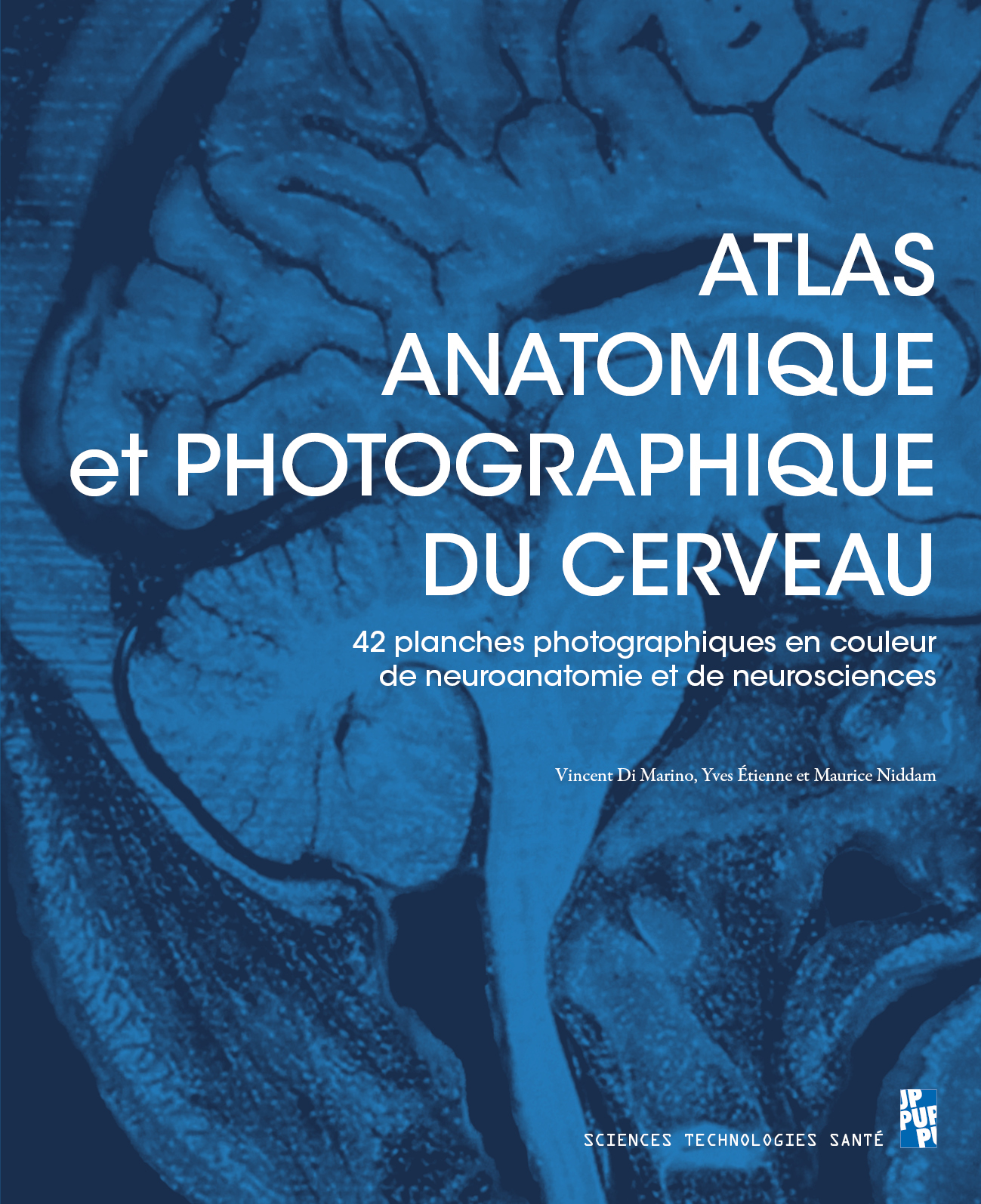
We didn't want to produce a traditional textbook, as there are already many available, most of them with beautiful captioned diagrams. We wanted to use photos to show future doctors the reality of the structures revealed by dissection. For the sections, too often of little interest because of their uniform pale hue, we benefited from a very interesting staining technique, discovered by chance in the anatomy laboratory. With Yves Etienne and Maurice Niddam, co-authors of the atlas, we had noticed that a brain sample which had been preserved in formalin (editor's note: use of formalin was still legal in the European Union at the time) and then immersed in a ferric chloride solution, took on a very interesting metallic hue, enhancing the structures and resulting in a perfect distinction between white and grey matter. The cross-sections shown in the atlas have been processed using this staining process, making our work even more special.
Throughout history, anatomy has often mingled with the artistic when it comes to representing the objects of study, particularly through drawing. Who produced the photographs and cross-sections for this book?
It's true that drawing enabled early anatomists to get closer to reality. An example of this is Foville's drawing of the cingulum fasciculus (1833), which is very similar to the photograph of dissection of the fasciculus in the atlas. But in this day and age, nothing is more demonstrative than photographs, especially if they are taken at the end of the dissection, by the person who has just carried it out. This way of proceeding enabled us to obtain very explicit photos, taken from the most suitable angles (which would not have been possible with a professional photographer, but without anatomical knowledge!). As for the cuts, they were made under our instructions, our direction and in our presence, by the excellent technicians of the anatomy laboratory, following the classic procedure on frozen heads.
You've written a number of books, but this is the first one you've produced in French. Why this change?
Our first book on the anatomy of the nervous system was published in French. Subsequently, however, as English became the scientific language not only in English-speaking countries, but throughout the world, we entrusted Springer-Verlag with the English-language editions of all our scientific books. In the case of the current atlas, the book had been deliberately prepared for a French edition, as we intended it, above all, for our dear students of medicine, neuroscience and all "neuro" specialties. But when we contacted our former publisher Springer, we were told that from now on they would only publish in English. So began a difficult search for a French publisher, most of whom were reluctant to commit to an atlas containing more than 100 photos, and therefore quite expensive to publish. We were delighted to obtain a "commitment to publish" from the Presses Universitaires de Provence, after a favourable opinion from the sapiteurs who examined our work, for which we thank them.
FSMPM_couverture_atlas
Vincent Di Marino, former professor emeritus of anatomy, was the director of the Anatomy Laboratory at the Marseille Medical Faculty from 2001 to 2012.
Reference: Di Marino, V., Etienne, Y., & Niddam, M. (2024). Anatomical and photographic atlas of the brain: 42 plates (including 41 photographs) of neuroanatomy and neuroscience. Published in April 2024 by Presses Universitaires de Provence.
Article published on June 21, 2024.
Interview by Fanny Trifilieff.
Discover also


Share on social media or